
Bestand:Bronchiolar epithelium 3 - SEM.jpg

Grootte van deze voorvertoning: 585 × 599 pixels. Andere resoluties: 234 × 240 pixels | 469 × 480 pixels | 750 × 768 pixels | 1.024 × 1.049 pixels.
Oorspronkelijk bestand (1.024 × 1.049 pixels, bestandsgrootte: 375 kB, MIME-type: image/jpeg)
| Dit is een bestand van Wikimedia Commons. Onderstaande beschrijving komt van de beschrijving van het bestand daar. |
Beschrijving
| Beschrijving |
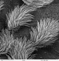
Scanning electron microscope image of lung trachea epithelium. There are both ciliated and non-ciliated cells in this epithelium. Note the difference in size between the cilia and the microvilli (on the non-ciliated cell surface). Zeiss DSM 962 SEM |
| Bron | |
| Auteur | Charles Daghlian |
| Toestemming (Hergebruik van dit bestand) |
PD |
Licentie
| Dit werk vrijgegeven in het publieke domein door de auteur, Charles Daghlian. Dit is wereldwijd van toepassing. In sommige landen is dit wettelijk niet mogelijk; in die gevallen geldt: Charles Daghlian staat iedereen toe dit werk voor eender welk doel te gebruiken, zonder enige voorwaarden, tenzij zulke voorwaarden door de wet worden voorgeschreven.
|
Bestandsgeschiedenis
Klik op een datum/tijd om het bestand te zien zoals het destijds was.
| Datum/tijd | Miniatuur | Afmetingen | Gebruiker | Opmerking | |
|---|---|---|---|---|---|
| huidige versie | 7 okt 2006 16:16 | | 1.024 × 1.049 (375 kB) | Patho | {{Information |Description=Scanning electron microscope image of lung trachea epithelium. There are both ciliated and on-ciliated cells in this epithelium. Note the difference in size between the cilia and the microvilli(on non-ciliated cell surface) Zei |
Bestandsgebruik
Dit bestand wordt op de volgende pagina gebruikt:
Globaal bestandsgebruik
De volgende andere wiki's gebruiken dit bestand:
- Gebruikt op ar.wikipedia.org
- Gebruikt op ast.wikipedia.org
- Gebruikt op bs.wikipedia.org
- Gebruikt op ca.wikipedia.org
- Gebruikt op cs.wikipedia.org
- Gebruikt op da.wikipedia.org
- Gebruikt op de.wikipedia.org
- Gebruikt op de.wikibooks.org
- Gebruikt op en.wikipedia.org
- Gebruikt op es.wikipedia.org
- Gebruikt op eu.wikipedia.org
- Gebruikt op fa.wikipedia.org
- Gebruikt op fr.wikipedia.org
- Gebruikt op gl.wikipedia.org
- Gebruikt op he.wikipedia.org
- Gebruikt op he.wiktionary.org
- Gebruikt op hi.wikipedia.org
- Gebruikt op id.wikipedia.org
- Gebruikt op jv.wikipedia.org
- Gebruikt op kk.wikipedia.org
- Gebruikt op lt.wikipedia.org
- Gebruikt op lv.wikipedia.org
- Gebruikt op ms.wikipedia.org
- Gebruikt op nn.wikipedia.org
- Gebruikt op no.wikipedia.org
- Gebruikt op pl.wikipedia.org
- Gebruikt op pl.wiktionary.org
- Gebruikt op pt.wikipedia.org
- Gebruikt op ru.wikipedia.org
- Gebruikt op ru.wiktionary.org
- Gebruikt op sh.wikipedia.org
- Gebruikt op simple.wikipedia.org
Globaal gebruik van dit bestand bekijken.