
Bestand:Echinococcus Life Cycle.png
Echinococcus_Life_Cycle.png (600 × 571 pixels, bestandsgrootte: 44 kB, MIME-type: image/png)
| Dit is een bestand van Wikimedia Commons. Onderstaande beschrijving komt van de beschrijving van het bestand daar. |
Beschrijving
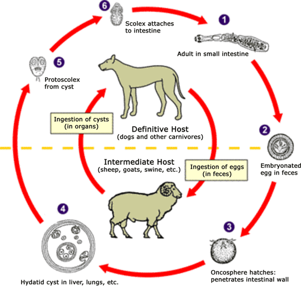
The adult Echinococcus granulosus (3 to 6 mm long) [1] resides in the small bowel of the definitive hosts (dogs or other carnivores). Gravid proglottids release eggs [2] that are passed in the feces. After ingestion by a suitable intermediate host (under natural conditions: sheep, goat, swine, cattle, horses, camel), the egg hatches in the small bowel and releases an oncosphere [3] that penetrates the intestinal wall and migrates through the circulatory system into various organs, especially the liver and lungs. In these organs, the oncosphere develops into a cyst [4] that enlarges gradually, producing protoscolices and daughter cysts that fill the cyst interior. The definitive host becomes infected by ingesting the cyst-containing organs of the infected intermediate host. After ingestion, the protoscolices [5] evaginate, attach to the intestinal mucosa [6] and develop into adult stages [1] in 32 to 80 days. The same life cycle occurs with E. multilocularis (1.2 to 3.7 mm), with the following differences: the definitive hosts are foxes, and to a lesser extent dogs, cats, coyotes and wolves; the intermediate host are small rodents; and larval growth (in the liver) remains indefinitely in the proliferative stage, resulting in invasion of the surrounding tissues. With E. vogeli (up to 5.6 mm long), the definitive hosts are bush dogs and dogs; the intermediate hosts are rodents; and the larval stage (in the liver, lungs and other organs) develops both externally and internally, resulting in multiple vesicles. E. oligarthrus (up to 2.9 mm long) has a life cycle that involves wild felids as definitive hosts and rodents as intermediate hosts. Humans become infected by ingesting eggs , with resulting release of oncospheres in the intestine and the development of cysts in various organs.
Image adapted from original available at the United States Centres for Disease Control Parasitology Identification Laboratory ([1] archief kopie op de Wayback Machine).

|
Bestand:Echinococcus Life Cycle.svg is een vectorversie van dit bestand. Indien niet van slechtere kwaliteit dient deze gebruikt te worden in plaats van deze rasterafbeelding.
File:Echinococcus Life Cycle.png → File:Echinococcus Life Cycle.svg
Zie Help:SVG voor meer informatie. |
 |
Licentie
Deze afbeelding is een werk van de Centers for Disease Control and Prevention, onderdeel van de United States Department of Health and Human Services, genomen of gemaakt tijdens de officiële werkzaamheden van een werknemer. Als werk van de Federale overheid van de Verenigde Staten, bevindt de afbeelding zich in het publiek domein.
|
Bestandsgeschiedenis
Klik op een datum/tijd om het bestand te zien zoals het destijds was.
| Datum/tijd | Miniatuur | Afmetingen | Gebruiker | Opmerking | |
|---|---|---|---|---|---|
| huidige versie | 24 jan 2007 12:54 | | 600 × 571 (44 kB) | Pngbot | optimized with optipng |
| 26 apr 2005 03:48 |  | 600 × 571 (55 kB) | FirstPrinciples~commonswiki | Smaller & clearer | |
| 26 apr 2005 03:36 |  | 800 × 761 (79 kB) | FirstPrinciples~commonswiki |
Bestandsgebruik
Geen enkele pagina gebruikt dit bestand.