
Bestand:Human brain anterior-inferior view description.JPG
Human_brain_anterior-inferior_view_description.JPG (330 × 475 pixels, bestandsgrootte: 31 kB, MIME-type: image/jpeg)
| Dit is een bestand van Wikimedia Commons. Onderstaande beschrijving komt van de beschrijving van het bestand daar. |
Beschrijving
| Beschrijving |
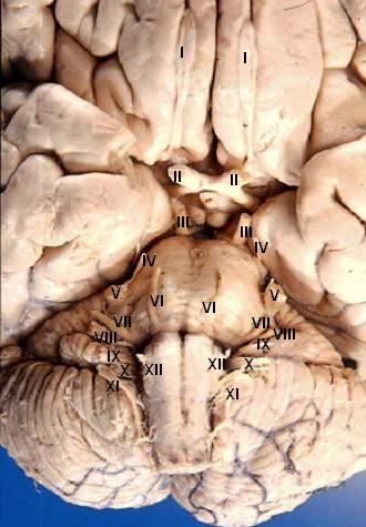
Human brain - anterior-inferior view - cerebral nerves I. N. olfactorius - The fila olfactoria (approximately 20 nerve rootlets on each side) comprise the Olfactory nerves (I) which run from the olfactory receptors in the nasal cavity to the olfactory bulb. II. N. opticus - The Optic nerves (II) are large & join at the midline to form the optic chiasm, then continue laterally as the optic tracts. The optic nerve is not a true nerve but rather a CNS tract. III. N. oculomotorius - The Oculomotor nerves (III) emerge from a depression in the midbrain, the interpeduncular fossa, just caudal to the optic chiasm. IV. N. trochlearis - The Trochlear nerves (IV) are small & are seen on the lateral surface of the midbrain. They are the only nerves which exit from the posterior side of the brain. V. N. trigeminus - The Trigeminal nerves (V) are large and emanate from the lateral surface of the pons. VI. N. abducens - The Abducens nerves (VI) exit near the midline from the inferior pontine sulcus which separates the pons from the medulla. VII. N. facialis - Moving laterally in the inferior pontine sulcus, the Facial nerves (VII) can be seen. VIII. N. vestibulocochlearis - Slightly lateral to the facial nerve is the Vestibulocochlear nerve (VIII). IX. N. glossopharyngeus - The postolivary sulcus is a groove running rostrocaudally on the lateral surface of the medulla. From this sulcus pass the small Glossopharyngeal nerves (IX) rostrally and... X. N. vagus - ...the much larger Vagus nerves (X) caudally. XI. N. accessorius - The Spinal Accessory nerves (XI) exit the cervical cord then pass rostrally through the foramen magnum to exit the cranial vault with the lossopharyngeal and vagus nerves. XII. N. hypoglossus - The Hypoglossal nerves (XII) exit the medulla via the preolivary sulcus.
|
| Datum | |
| Bron | http://www.healcentral.org/healapp/showMetadata?metadataId=40566 (Internet Archive of file description page) |
| Auteur |
John A Beal, PhD Dep't. of Cellular Biology & Anatomy, Louisiana State University Health Sciences Center Shreveport |
| Toestemming (Hergebruik van dit bestand) |
CC-BY |
| Andere versies |
 |
Licentie
- De gebruiker mag:
- Delen – het werk kopiëren, verspreiden en doorgeven
- Remixen – afgeleide werken maken
- Onder de volgende voorwaarden:
- naamsvermelding – U moet op een gepaste manier aan naamsvermelding doen, een link naar de licentie geven, en aangeven of er wijzigingen in het werk zijn aangebracht. U mag dit op elke redelijke manier doen, maar niet zodanig dat de indruk wordt gewekt dat de licentiegever instemt met uw werk of uw gebruik van zijn werk.
Dit bestand, dat oorspronkelijk toegevoegd was op http://www.healcentral.org/healapp/showMetadata?metadataId=40566, werd op 25 september 2013 beoordeeld door de moderator of reviewer Eleassar, die bevestigt dat dit bestand beschikbaar was onder de vermelde licentie op die datum.
|
Bestandsgeschiedenis
Klik op een datum/tijd om het bestand te zien zoals het destijds was.
| Datum/tijd | Miniatuur | Afmetingen | Gebruiker | Opmerking | |
|---|---|---|---|---|---|
| huidige versie | 24 jun 2006 20:22 | | 330 × 475 (31 kB) | Patho | {{Information| |Description='''Human brain - anterior-inferior view - cerebral nerves''' I. N. olfactorius - The fila olfactoria (approximately 20 nerve rootlets on each side) comprise the Olfactory nerves (I) which run from the olfactory receptors in |
Bestandsgebruik
Dit bestand wordt op de volgende pagina gebruikt:
Globaal bestandsgebruik
De volgende andere wiki's gebruiken dit bestand:
- Gebruikt op ar.wikipedia.org
- Gebruikt op azb.wikipedia.org
- Gebruikt op az.wikipedia.org
- Gebruikt op bs.wikipedia.org
- Gebruikt op de.wikipedia.org
- Gebruikt op de.wikibooks.org
- Gebruikt op en.wikipedia.org
- Cranial nerves
- User talk:Hovea
- User talk:Wouterstomp
- User talk:Nephron
- User talk:NCurse
- User talk:Robotsintrouble
- User talk:Was a bee
- User talk:Bloomingdedalus
- User talk:Read-write-services
- User talk:Attys
- User talk:Bakerstmd
- User talk:Manfi
- User talk:Mikepascoe
- User talk:Vokesk
- User talk:Neuraxıs
- User talk:Cmungall
- User talk:Qxukhgiels
- User talk:DocElisa
- User talk:JakobSteenberg/Archives/1
- User talk:Meteor sandwich yum
- User talk:Slashedone
- User talk:Anindya07
- User talk:Mattimussi
- User talk:Jelly Bean MD
- User talk:Benrusholme
- User talk:Brad.w.english
- User talk:Krsna ss
- User talk:Tom (LT)/Archive 4
- User talk:Dregon131
- User talk:Sapientia42
- User talk:Tilifa Ocaufa
- User talk:Snow Rise/Archive 6
- User talk:Arcadian
- User talk:Outofbattery
- User talk:Anatomyczar
- Wikipedia:WikiProject Anatomy/Newsletter/4
- User talk:Supravibhatsupravi
- User talk:Athikhun.suw
- User talk:CFCF/Archive 6
- User talk:Lewisskinner/ArchiveJul 2015
- User talk:Tyrol5/Archive 6
- User talk:CFCF/Archive 7
- User talk:DiverDave/Archive 5
Globaal gebruik van dit bestand bekijken.