
Bestand:Paracentrotus lividus skeletogenesis.jpg

Grootte van deze voorvertoning: 445 × 600 pixels. Andere resoluties: 178 × 240 pixels | 356 × 480 pixels | 570 × 768 pixels | 1.243 × 1.675 pixels.
Oorspronkelijk bestand (1.243 × 1.675 pixels, bestandsgrootte: 1,63 MB, MIME-type: image/jpeg)
| Dit is een bestand van Wikimedia Commons. Onderstaande beschrijving komt van de beschrijving van het bestand daar. |
Beschrijving
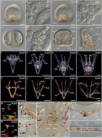
| Beschrijving | FIGURE 9. Skeletogenesis in Paracentrotus lividus during the embryonic and larval periods. Developmental stages are as follows: (A,B) early gastrula stage (EG); (C,D) mid-gastrula stage (mid-G); (E,F) late gastrula stage (LG); (G,H) prism stage (prism); (I,I') 2-arm pluteus stage (2-arm); (J,J′) 4-arm pluteus stage (4-arm); (K,K',M–P) 6-arm pluteus stage (6-arm); (L,L',Q–S) 8-arm pluteus stage (8-arm). In (A–L,N,P–S), images were acquired using light microscopy either in bright-field for (A–H,N,P–S), or dark-field for (I–L). In (I'–L',M,O), images were obtained using polarized light to highlight the skeletal elements. In (A,C,H), embryos are in right view, with the animal pole up and, for (H), with the ventral side left. In (E,G), embryos are in ventral view, with the animal pole up. In (I–L′), larvae are in anterior view, with the ventral side up. (B) Close-up of the skeletogenic mesoderm cells and their related filopodia in a ventral chain. (D,F) Close-ups of a ventrolateral cluster to highlight the rhombohedral crystal in (D) and the triradiate spicule in (F), the two images corresponding, respectively, to the regions highlighted by yellow boxes in (C) and (E). (M–P) Close-ups of the regions outlined by orange boxes in (K) with larvae in lateral view, except for (M) where the larva is in anterior view. (M,N) Close-ups of the developing posterodorsal spicule. (O,P) Close-ups of the developing dorsal arch. (Q–S) Close-ups of the regions highlighted by orange boxes in (L) with larvae in anterior view. (Q) Close-up of the skeletal elements located in the vicinity of the larval digestive tract. (R) Close-up of the body rods in the most dorsal region of the larva. (S) Close-up of the non-fenestrated spicule of an anterolateral arm. In (B), arrowheads highlight the filopodia extended by skeletogenic mesoderm cells. In (C–F), double arrowheads mark the skeletal elements forming in the ventrolateral clusters. In (H), the arrowhead highlights the limit between the right dorsoventral connecting rod and the right anterolateral rod. In (I,J), the asterisk marks the oral hood. Scale bar: (A,C,E,G,H,M–S) 30 μm; (B,D,F) 15 μm; (I) 100 μm; (J–L,J′–L′) 200 µm. AmR: anteromedial rod; Apx: apex; DA: dorsal arch; lAdtR: left anterodorsal transverse rod; lAla: left anterolateral arm; lAlR: left anterolateral rod; lAvtR: left anteroventral transverse rod; lBR: left body rod; lDvcR: left dorsoventral connecting rod; lPoa: left postoral arm; lPoR: left postoral rod; lPda: left posterodorsal arm; lPdR: left posterodorsal rod; lPra: left preoral arm; lPrR: left preoral rod; lRR: left recurrent rod; lVtR: left ventral transverse rod; rAdtR: right anterodorsal transverse rod; rAla: right anterolateral arm; rAlR: right anterolateral rod; rAvtR: right anteroventral transverse rod; rBR: right body rod; rDvcR: right dorsoventral connecting rod; rPoa: right postoral arm; rPoR: right postoral rod; rPda: right posterodorsal arm; rPdR: right posterodorsal rod; rPra: right preoral arm; rPrR: right preoral rod; rRR: right recurrent rod; rVLC: right ventrolateral cluster; rVtR: right ventral transverse rod; St: stomach. |
| Datum | |
| Bron |
https://www.frontiersin.org/articles/10.3389/fcell.2022.966408/full Developmental atlas of the indirect-developing sea urchin Paracentrotus lividus: From fertilization to juvenile stages, Front. Cell Dev. Biol., 31 October 2022 Sec. Morphogenesis and Patterning Volume 10 - 2022, https://doi.org/10.3389/fcell.2022.966408 |
| Auteur | Laurent Formery, Axel Wakefield, Maeva Gesson, Ludovic Toisoul, Guy Lhomond, Laurent Gilletta, Régis Lasbleiz, Michael Schubert, Jenifer C. Croce1 |
Licentie
Dit bestand is gelicenseerd onder de Creative Commons Naamsvermelding 4.0 Internationaal licentie.
- De gebruiker mag:
- Delen – het werk kopiëren, verspreiden en doorgeven
- Remixen – afgeleide werken maken
- Onder de volgende voorwaarden:
- naamsvermelding – U moet op een gepaste manier aan naamsvermelding doen, een link naar de licentie geven, en aangeven of er wijzigingen in het werk zijn aangebracht. U mag dit op elke redelijke manier doen, maar niet zodanig dat de indruk wordt gewekt dat de licentiegever instemt met uw werk of uw gebruik van zijn werk.

|
Dit bestand, dat oorspronkelijk toegevoegd was op een externe website, is nog niet beoordeeld door een moderator of reviewer om te bevestigen dat de opgegeven licentie geldig is. Zie Category:License review needed voor meer instructies.
|
Bestandsgeschiedenis
Klik op een datum/tijd om het bestand te zien zoals het destijds was.
| Datum/tijd | Miniatuur | Afmetingen | Gebruiker | Opmerking | |
|---|---|---|---|---|---|
| huidige versie | 6 mrt 2024 00:25 | | 1.243 × 1.675 (1,63 MB) | Rasbak | {{Information |description=FIGURE 9. Skeletogenesis in Paracentrotus lividus during the embryonic and larval periods. Developmental stages are as follows: (A,B) early gastrula stage (EG); (C,D) mid-gastrula stage (mid-G); (E,F) late gastrula stage (LG); (G,H) prism stage (prism); (I,I') 2-arm pluteus stage (2-arm); (J,J′) 4-arm pluteus stage (4-arm); (K,K',M–P) 6-arm pluteus stage (6-arm); (L,L',Q–S) 8-arm pluteus stage (8-arm). In (A–L,N,P–S), images were acquired using light microscopy eith... |
Bestandsgebruik
Dit bestand wordt op de volgende pagina gebruikt: