
Bestand:Transver cells fpls-05-00046-g006.jpg

Grootte van deze voorvertoning: 495 × 599 pixels. Andere resoluties: 198 × 240 pixels | 396 × 480 pixels | 645 × 781 pixels.
Oorspronkelijk bestand (645 × 781 pixels, bestandsgrootte: 389 kB, MIME-type: image/jpeg)
| Dit is een bestand van Wikimedia Commons. Onderstaande beschrijving komt van de beschrijving van het bestand daar. |
Beschrijving
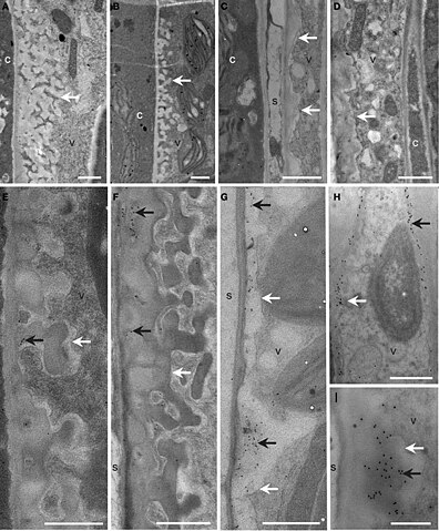
| Beschrijving | Figure 6. Cellular structure and immunodetection of callose after 3 days of LT treatment. Col, gsl5, vte2, and gsl5 vte2 were grown under permissive conditions for 4 weeks and transferred to LT conditions for 3 additional days. Col (A,E), gsl5 (B,F), vte2 (C,G), and gsl5 vte2 (D,H,I). Black arrows highlight wall ingrowths of phloem parenchyma transfer cells immunolabeled with anti-β-1,3-glucan. White arrows mark transfer cell walls. c, companion cell; s, sieve element; v, vascular parenchyma transfer cell. Bars = 1 μm (A–H), 0.5 μm (I). |
| Datum | |
| Bron | https://www.frontiersin.org/files/Articles/76650/fpls-05-00046-HTML/image_m/fpls-05-00046-g006.jpg https://www.frontiersin.org/articles/10.3389/fpls.2014.00046/full Role of callose synthases in transfer cell wall development in tocopherol deficient Arabidopsis mutants, Front. Plant Sci., 19 February 2014, Sec. Plant Physiology, Volume 5 - 2014 |
| Auteur | Hiroshi Maeda1, Wan Song1, Tammy Sage, Dean DellaPenna1 |
- Error in {{Information}} template: unknown parameter "1".
Open access
Licentie
Dit bestand is gelicenseerd onder de Creative Commons-licentie Naamsvermelding 3.0 Unported
- De gebruiker mag:
- Delen – het werk kopiëren, verspreiden en doorgeven
- Remixen – afgeleide werken maken
- Onder de volgende voorwaarden:
- naamsvermelding – U moet op een gepaste manier aan naamsvermelding doen, een link naar de licentie geven, en aangeven of er wijzigingen in het werk zijn aangebracht. U mag dit op elke redelijke manier doen, maar niet zodanig dat de indruk wordt gewekt dat de licentiegever instemt met uw werk of uw gebruik van zijn werk.
Bestandsgeschiedenis
Klik op een datum/tijd om het bestand te zien zoals het destijds was.
| Datum/tijd | Miniatuur | Afmetingen | Gebruiker | Opmerking | |
|---|---|---|---|---|---|
| huidige versie | 1 jan 2024 20:23 | | 645 × 781 (389 kB) | Rasbak | {{Information |description=Figure 6. Cellular structure and immunodetection of callose after 3 days of LT treatment. Col, gsl5, vte2, and gsl5 vte2 were grown under permissive conditions for 4 weeks and transferred to LT conditions for 3 additional days. Col (A,E), gsl5 (B,F), vte2 (C,G), and gsl5 vte2 (D,H,I). Black arrows highlight wall ingrowths of phloem parenchyma transfer cells immunolabeled with anti-β-1,3-glucan. White arrows mark transfer cell walls. c, companion cell; s, sieve eleme... |
Bestandsgebruik
Dit bestand wordt op de volgende pagina gebruikt: