
Bestand:Abatus cordatus Segmentation.jpg

Oorspronkelijk bestand (1.640 × 1.592 pixels, bestandsgrootte: 665 kB, MIME-type: image/jpeg)
| Dit is een bestand van Wikimedia Commons. Onderstaande beschrijving komt van de beschrijving van het bestand daar. |
Beschrijving
| Beschrijving |
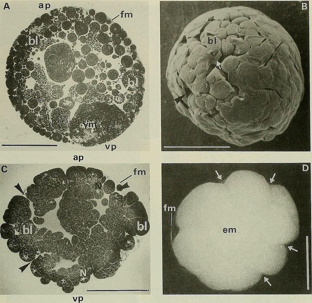
Figure 4. Segmentation of Abatus cordatus. (A) Section through a cleaved egg showing blastomeres and the remaining yolk mass at the vegetal pole (11 days after fertilization). (B) SEM view of a completely cleaved egg (14 days after fertilization). Blastomeres are visible where the fertilization membrane is destroyed (white arrow). Black arrow shows a furrow. (C) Section through a cleaved egg at the end of the segmentation (15 days after fertilization). Black arrows show the large intercellular spaces that communicate with the perivitelline space at the level of the furrows. (D) Light micrograph of a wrinkled stereoblastula (26 days after fertilization). Arrows show furrows. A and C are semi-thin plastic sections (toluidine blue, pH 11.5). Abbreviations: ap, animal pole; bl, blastomere; em, embryo; fm, fertilization membrane; N, nuclear area; vp, vegetal pole; ym, yolk mass. Scale bar = 500 um |
| Datum | |
| Bron | https://www.flickr.com/photos/internetarchivebookimages/20191999800/ The Biological bulletin. Marine Biological Laboratory (Woods Hole, Mass. ). Annual report; HighWire Press. |
| Auteur | Lillie, Frank Rattray, Moore, Carl Richard, Redfield, Alfred Clarence. |
Licentie
| Dit bestand is beschikbaar onder Creative Commons CC0 1.0 Universele Public Domain Dedication. | |
| De persoon die een werk voorziet van deze licentie stelt dit werk beschikbaar aan het publieke domein door, voor zover dit wettelijk is toegestaan, afstand te doen van alle rechten op het werk in de zin van het auteursrecht, met inbegrip van alle aanverwante of naburige rechten. U kunt het werk kopiëren, aanpassen, distribueren en uitvoeren, ook voor commerciële doeleinden, zonder dat u daarvoor toestemming hoeft te vragen.
|
| Deze afbeelding is oorspronkelijk op Flickr geplaatst door Internet Archive Book Images op https://flickr.com/photos/126377022@N07/20191999800. Dit is op 25 maart 2024 door de FlickreviewR 2-Bot beoordeeld en de licentie onder de voorwaarden van cc-zero is bevestigd. |
Bestandsgeschiedenis
Klik op een datum/tijd om het bestand te zien zoals het destijds was.
| Datum/tijd | Miniatuur | Afmetingen | Gebruiker | Opmerking | |
|---|---|---|---|---|---|
| huidige versie | 25 mrt 2024 13:36 | | 1.640 × 1.592 (665 kB) | Rasbak | {{information |description= Figure 4. Segmentation of Abatus cordatus. (A) Section through a cleaved egg showing blastomeres and the remaining yolk mass at the vegetal pole (11 days after fertilization). (B) SEM view of a completely cleaved egg (14 days after fertilization). Blastomeres are visible where the fertilization membrane is destroyed (white arrow). Black arrow shows a furrow. (C) Section through a cleaved egg at the end of the segmentation (15 days after fertilization). Black arrows... |
Bestandsgebruik
Dit bestand wordt op de volgende pagina gebruikt: