
Bestand:Brain - Lobes.png
Geen hogere resolutie beschikbaar.
Brain_-_Lobes.png (701 × 487 pixels, bestandsgrootte: 360 kB, MIME-type: image/png)
| Dit is een bestand van Wikimedia Commons. Onderstaande beschrijving komt van de beschrijving van het bestand daar. |
| Beschrijving |
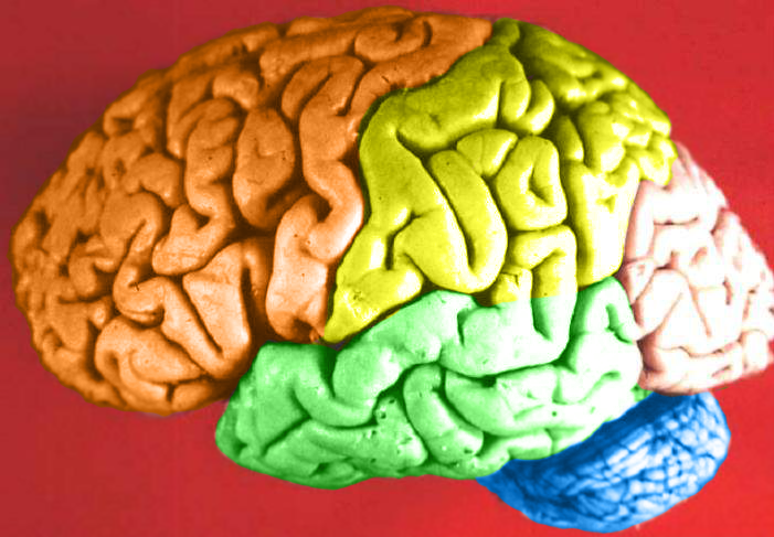
Human brain lateral view - Lobes
|
| Datum | (UTC) |
| Bron | Human_brain_lateral_view_description.JPG |
| Auteur | Dep't. of Cellular Biology & Anatomy, Louisiana State University Health Sciences Center Shreveport |
| Toestemming (Hergebruik van dit bestand) |
CC-BY |
| Andere versies |
| Dit is een geretoucheerde foto, wat betekent dat de originele versie digitaal aangepast is. Aanpassingen: Hemispheres in color.. Het origineel kan hier bekeken worden: Human brain lateral view description.JPG. Aanpassingen gedaan door DavoO.
|
Licentie
Ik, de auteursrechthebbende van dit werk, maak het hierbij onder de volgende licentie beschikbaar:
Dit bestand is gelicenseerd onder de Creative Commons-licentie Naamsvermelding 2.5 Unported
- De gebruiker mag:
- Delen – het werk kopiëren, verspreiden en doorgeven
- Remixen – afgeleide werken maken
- Onder de volgende voorwaarden:
- naamsvermelding – U moet op een gepaste manier aan naamsvermelding doen, een link naar de licentie geven, en aangeven of er wijzigingen in het werk zijn aangebracht. U mag dit op elke redelijke manier doen, maar niet zodanig dat de indruk wordt gewekt dat de licentiegever instemt met uw werk of uw gebruik van zijn werk.
The following refers to the original source file, not this derivative version.
Dit bestand, dat oorspronkelijk toegevoegd was op https://web.archive.org/web/20110514023714/http://www.healcentral.org/healapp/showMetadata?metadataId=40566, werd op 1 november 2013 beoordeeld door de moderator of reviewer Avenue, die bevestigt dat dit bestand beschikbaar was onder de vermelde licentie op die datum.
|
Oorspronkelijk uploadlogboek
This image is a derivative work of the following images:
- File:Human_brain_lateral_view_description.JPG licensed with Cc-by-2.5
- 2006-06-20T13:58:22Z Patho 701x487 (50176 Bytes) {{Information| |Description='''Human brain lateral view - Lobes''' # Lobus frontalis # Lobus parietalis # Lobus temporalis # Lobus occipitalis # Sulcus lateralis # Sulcus centralis # Sulcus parietooccipitalis # Incisura preo
- 2006-06-20T13:54:13Z Patho 701x487 (49891 Bytes) Auf eine alte Version zurückgesetzt
- 2006-06-20T13:51:38Z Patho 701x487 (50074 Bytes) {{Information| |Description='''Human brain lateral view - Lobes''' # Lobus frontalis # Lobus parietalis # Lobus temporalis # Lobus occipitalis # Sulcus lateralis # Sulcus centralis # Sulcus parietooccipitalis # Incisura preo
- 2006-06-20T13:28:44Z Patho 701x487 (49891 Bytes) {{Information| |Description='''Human brain lateral view''' # Lobus frontalis # Lobus parietalis # Lobus temporalis # Lobus occipitalis # sulcus lateralis # Sulcis centralis # Sulcus parietooccipitalis # Incisura preoccipital
Uploaded with derivativeFX
Bestandsgeschiedenis
Klik op een datum/tijd om het bestand te zien zoals het destijds was.
| Datum/tijd | Miniatuur | Afmetingen | Gebruiker | Opmerking | |
|---|---|---|---|---|---|
| huidige versie | 24 feb 2009 00:11 | | 701 × 487 (360 kB) | DavoO | {{Information |Description='''Human brain lateral view - Lobes''' # Lobus frontalis # Lobus parietalis # Lobus temporalis # Lobus occipitalis # Sulcus lateralis # Sulcus centralis # Sulcus parietooccipitalis # Incisura preoccipitalis # Polus frontalis # |
Bestandsgebruik
Dit bestand wordt op de volgende pagina gebruikt:
Globaal bestandsgebruik
De volgende andere wiki's gebruiken dit bestand:
- Gebruikt op cs.wikipedia.org
- Gebruikt op en.wikipedia.org
- Talk:Alcohol intoxication
- Talk:LSD
- Talk:Scopolamine
- Talk:Qigong
- Talk:Recreational drug use
- Talk:Psilocybin
- Talk:Phenomenology (philosophy)
- Talk:Alcohol (chemistry)
- Talk:Timothy Leary
- Talk:Psilocybe cubensis
- Talk:Nitrous oxide
- Talk:Atropine
- Talk:Out-of-body experience
- Talk:The Doors of Perception
- Talk:Carlos Castaneda
- Talk:Ganzfeld experiment
- Talk:Meditation
- Talk:Zen
- Talk:Hysteria
- Talk:Peyote
- Talk:Hashish
- Talk:Ketamine
- Talk:Coca
- Talk:Hippie
- Talk:Spirituality
- Talk:Mantra
- Talk:N,N-Dimethyltryptamine
- Talk:Amanita muscaria
- Talk:Hookah
- Talk:Autogenic training
- Talk:Psychonautics
- Talk:Hypnosis
- Talk:Dipropyltryptamine
- Talk:Ergot
- Talk:Phencyclidine
- Talk:Anadenanthera peregrina
- Talk:Shamanism
- Talk:Mescaline
- Talk:Diphenhydramine
- Talk:Salvinorin A
- Talk:Human Potential Movement
- Talk:Argyreia nervosa
- Talk:Terence McKenna
- Talk:Psilocybin mushroom
- Talk:Atropa belladonna
- Talk:Hypnagogia
- Talk:Kundalini yoga
- Talk:DiPT
- Talk:Dreamachine
Globaal gebruik van dit bestand bekijken.