
Bestand:Paracentrotus lividus anatomy of early and late juveniles.jpg

Grootte van deze voorvertoning: 533 × 600 pixels. Andere resoluties: 213 × 240 pixels | 426 × 480 pixels | 682 × 768 pixels | 1.117 × 1.257 pixels.
Oorspronkelijk bestand (1.117 × 1.257 pixels, bestandsgrootte: 2,01 MB, MIME-type: image/jpeg)
| Dit is een bestand van Wikimedia Commons. Onderstaande beschrijving komt van de beschrijving van het bestand daar. |
Beschrijving
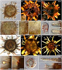
| Beschrijving | FIGURE 16. Anatomy of early and late juveniles of Paracentrotus lividus. Developmental stages are as follows: (A–I) early juvenile stage, 1 day post-metamorphosis (EJuv); (J–Q) late juvenile stage, 8 days post-metamorphosis (LJuv). In (A,D–J,M–Q), images were acquired using bright-field light microscopy. In (B,C,K,L), images were obtained using polarized light microscopy. In (A–D,J–L), images correspond to specimens in oral view in (A,C,D,J,L) and in aboral view in (B,K). ((B) inset) Close-up of an opened pedicellaria. ((C) inset) Close-up of the skeletal elements constituting the primordia of the adult masticatory apparatus. (D) Close-up of the ring and radial canals of the water vascular system. (E) Close-up of a pair of growing secondary podia located underneath a primary podium. (F) Close-up of the ampullae formed at the proximal tip of a radial canal. (G) Close-up of the skeletal disk within the papilla of a primary podium. (H) Close-up of a developing definitive spine. (I) Close-up of a developing juvenile spine. (M) Close-up of a sphaeridium. (N) Close-up of a degenerated primary podium. (O) Close-up of the skeletal disk within the papilla of a secondary podium. (P) Close-up of a fully formed definitive spine. (Q) Close-up of a fully formed juvenile spine. In (B), cyan asterisks mark the pedicellariae present on the aboral surface of the juvenile, and white dotted lines delineate the genital plates. In ((C) inset), the white arrowhead indicates a tooth primordium, cyan arrowheads highlight primordia of the hemipyramids, and green arrowheads mark primordia of the epiphyses. In (F), white dotted lines outline two ampullae formed at the proximal tip of a radial canal. In (H,I), white arrowheads indicate the apex of a growing definitive spine in (H) and of a growing juvenile spine in (I). Scale bar: (A–C,J–L) 100 μm; ((B) inset, (C) inset, (D–I,M–Q)) 30 µm. Amp: ampulla; AmbP: ambulacral plate; BucP: buccal plate; DS: definitive spine; gAB: genital plate AB; gBC: genital plate BC; gCD: genital plate CD; gDE: genital plate DE; gEA: genital plate EA; JS: juvenile spine; Peri: peristome; PP: primary podium; RaC: radial canal; RiC: ring canal; SP: secondary podium; Sph: sphaeridium; SR: skeletal rod; Tub: tubercule. |
| Datum | |
| Bron |
https://www.frontiersin.org/articles/10.3389/fcell.2022.966408/full Developmental atlas of the indirect-developing sea urchin Paracentrotus lividus: From fertilization to juvenile stages, Front. Cell Dev. Biol., 31 October 2022 Sec. Morphogenesis and Patterning Volume 10 - 2022, https://doi.org/10.3389/fcell.2022.966408 |
| Auteur | Laurent Formery, Axel Wakefield, Maeva Gesson, Ludovic Toisoul, Guy Lhomond, Laurent Gilletta, Régis Lasbleiz, Michael Schubert, Jenifer C. Croce |
Licentie
Dit bestand is gelicenseerd onder de Creative Commons Naamsvermelding 4.0 Internationaal licentie.
- De gebruiker mag:
- Delen – het werk kopiëren, verspreiden en doorgeven
- Remixen – afgeleide werken maken
- Onder de volgende voorwaarden:
- naamsvermelding – U moet op een gepaste manier aan naamsvermelding doen, een link naar de licentie geven, en aangeven of er wijzigingen in het werk zijn aangebracht. U mag dit op elke redelijke manier doen, maar niet zodanig dat de indruk wordt gewekt dat de licentiegever instemt met uw werk of uw gebruik van zijn werk.

|
Dit bestand, dat oorspronkelijk toegevoegd was op een externe website, is nog niet beoordeeld door een moderator of reviewer om te bevestigen dat de opgegeven licentie geldig is. Zie Category:License review needed voor meer instructies.
|
Bestandsgeschiedenis
Klik op een datum/tijd om het bestand te zien zoals het destijds was.
| Datum/tijd | Miniatuur | Afmetingen | Gebruiker | Opmerking | |
|---|---|---|---|---|---|
| huidige versie | 7 mrt 2024 12:13 | | 1.117 × 1.257 (2,01 MB) | Rasbak | {{Information |description=FIGURE 16. Anatomy of early and late juveniles of Paracentrotus lividus. Developmental stages are as follows: (A–I) early juvenile stage, 1 day post-metamorphosis (EJuv); (J–Q) late juvenile stage, 8 days post-metamorphosis (LJuv). In (A,D–J,M–Q), images were acquired using bright-field light microscopy. In (B,C,K,L), images were obtained using polarized light microscopy. In (A–D,J–L), images correspond to specimens in oral view in (A,C,D,J,L) and in aboral view in... |
Bestandsgebruik
Dit bestand wordt op de volgende pagina gebruikt: