Bestand:2908 Germ Layers-02-nltxt.jpg

Pagina-inhoud wordt niet ondersteund in andere talen.
Uit Wikipedia, de vrije encyclopedie

Oorspronkelijk bestand(1.854 × 1.658 pixels, bestandsgrootte: 1,45 MB, MIME-type: image/jpeg)


Beschrijving

Beschrijving

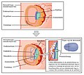
This image shows the different germ layers. The top panel shows the epiblast and trophoblast cells in the early stages of development. The bottom panel shows the three germ layers: the endoderm, ectoderm, and mesoderm. All the other major parts are also labeled. NLtxt labels.

Figure 28.9 Germ Layers Formation of the three primary germ layers occurs during the first 2 weeks of development. The embryo at this stage is only a few millimeters in length. Illustration from Anatomy & Physiology, Connexions Web site. Embryonic Development https://openstax.org/books/anatomy-and-physiology/pages/28-2-embryonic-development, Jun 19, 2013.
Datum
Bron Anatomy & Physiology, Connexions Web site. Embryonic Development https://openstax.org/books/anatomy-and-physiology/pages/28-2-embryonic-development, Jun 19, 2013.
Auteur OpenStax College
Andere versies

Licentie

w:nl:Creative Commons
naamsvermelding
Dit bestand is gelicenseerd onder de Creative Commons-licentie Naamsvermelding 3.0 Unported
De gebruiker mag:
  • Delen – het werk kopiëren, verspreiden en doorgeven
  • Remixen – afgeleide werken maken
Onder de volgende voorwaarden:
  • naamsvermelding – U moet op een gepaste manier aan naamsvermelding doen, een link naar de licentie geven, en aangeven of er wijzigingen in het werk zijn aangebracht. U mag dit op elke redelijke manier doen, maar niet zodanig dat de indruk wordt gewekt dat de licentiegever instemt met uw werk of uw gebruik van zijn werk.

Bijschriften

Beschrijf in één regel wat dit bestand voorstelt

Items getoond in dit bestand

beeldt af

image/jpeg

Bestandsgeschiedenis

Klik op een datum/tijd om het bestand te zien zoals het destijds was.

Datum/tijdMiniatuurAfmetingenGebruikerOpmerking
huidige versie22 feb 2024 23:57Miniatuurafbeelding voor de versie van 22 feb 2024 23:571.854 × 1.658 (1,45 MB)Rasbak{{Information |Description= This image shows the different germ layers. The top panel shows the epiblast and trophoblast cells in the early stages of development. The bottom panel shows the three germ layers: the endoderm, ectoderm, and mesoderm. All the other major parts are also labeled. Figure 28.9 Germ Layers Formation of the three primary germ layers occurs during the first 2 weeks of development. The embryo at this stage is only a few millimeters in length. Illustration from Anatomy &...

Dit bestand wordt op de volgende 6 pagina's gebruikt: